

The Program in Neuroscience provides lab and research opportunities in each of the core dimensions of our field:
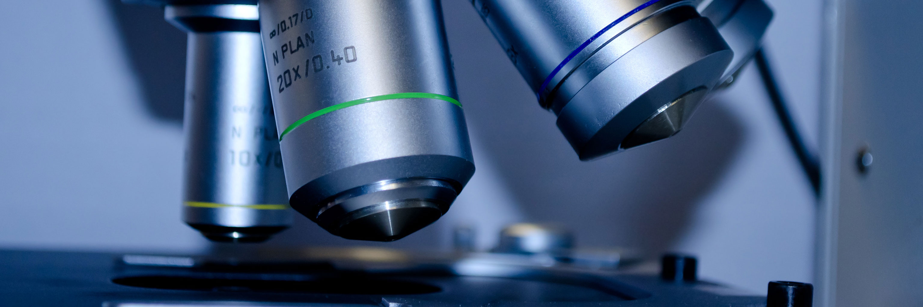
Image Research Facility
Molecular and Cellular Neuroscience
Behavioral Neuroscience
Cognitive and Computational Neuroscience
Clinical and Translational Neuroscience
Imaging Research Facility
The Imaging Research Facility (IRF) conducts research, training, and education in the study of brain structure and function and how these relate to behavior. Using multiple methods of neuro-imaging, it serves an interactive and collaborative community of researchers from across the IUB campus and beyond.
The IRF is extensively supported by the Department of Psychological and Brain Sciences and the College of Arts and Sciences.
MRI Technology
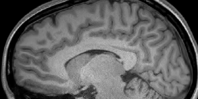
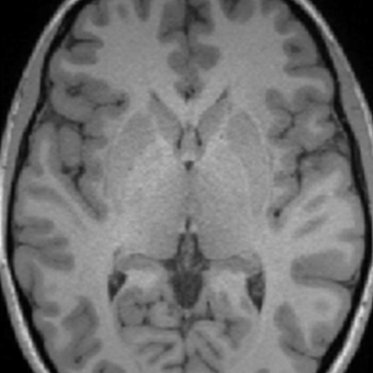
The IRF houses a Siemens 3 Tesla TIM Trio MRI scanner. Capabilities include structural MRI, functional MRI (fMRI), diffusion and perfusion imaging, and MR spectroscopy. Active tasks can be performed within the scanner using auditory, visual, and tactiledelivery methods. Participant responses such as eye blink, skin conductance, heart rate,and respiration have also been recorded during imaging.
High Resolution Anatomical Imaging
The IRF houses a whole body high field scanner capable of acquiring high-resolution images of any body part, including the brain and spinal cord. These high resolution images can be used in conjunction with both EEG and transcranial magnetic stimulation (TMS).
Functional Imaging
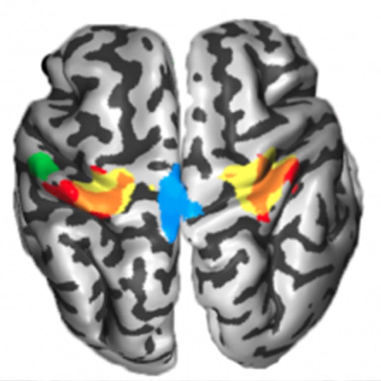
fMRI measures brain activity via oxygen metabolism. This technique, in conjunction with carefully designed behavioral tasks allows for the examination of brain functioning. There are projects exploring object recognition, child development, language processing, problem-solving, cognitive control, among others in the IRF. Additionally research is underway to explore brain functioning differences in clinical populations including schizophrenics, drug abusers and individuals with concussions.
Diffusion Imaging
Understanding how the brain is wired helps explain how it functions and how it learns. The white matter (the axons that connect neurons) allows for communication between neurons. By measuring signal change from the diffusion of water molecules, diffusion imaging can characterize the integrity of white matter and map the white matter pathways.
A number of clinical disorders have been shown to be correlated with white matter deficits, including cardiovascular disease and developmental disorders such as dyslexia and autism. A number of researchers in the IRF use this technology to explore drug abuse, schizophrenia and child development.
Perfusion Imaging
Perfusion imaging allows for the measure of cerebral blood flow and can be useful is the study of cardiovascular disease, stroke, and dementia.
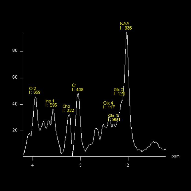
MR Spectroscopy
MRS allows for the in vivo quantification of neurotransmitters, such as GABA, as well as other substances, including manganese and iron. These measures can be acquired for specific brain regions. The ability to measure such substances allows for the investigation of differences in the concentration of various neurotransmitters as well as the study of neurotoxicity.
EEG Technology
EEG Capability
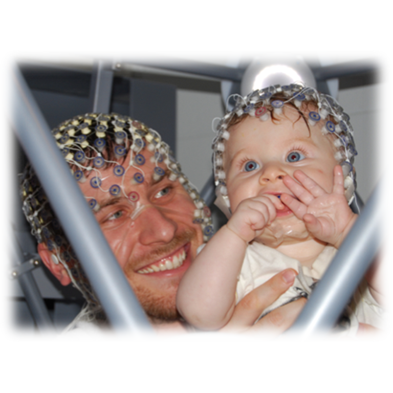
The IRF houses a laboratory which has a yoked and synchronized dual 64 channel EGI EEG system which allows for hyper-scanning of pairs of research participants during the execution of perceptual, cognitive or affective activation tasks with very high temporal resolution (to 1 ms accuracy).
The EEG system is also yoked to a Tobi infra-red eye tracking system which can be used in a stand-alone mode or in conjunction with the EEG system. The EEG system can be used to study special populations, including infants.
The ongoing EEG consists of a continuous varying signal composed of various frequencies that change with the subject’s state of alertness and perceptual, cognitive, and affective demands, allowing for the performance of a number of analyses.
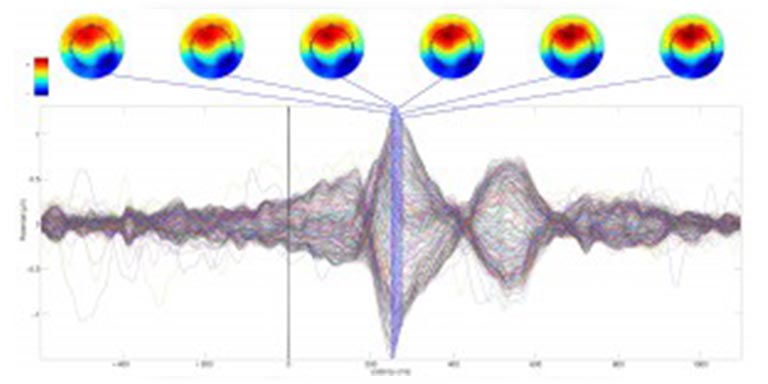
Event-Related Potentials
By synching stimulus delivery with EEG data acquisition, stimulus-locked activity (event-related potentials) can be recorded. Analysis in the time domain of peak ERP amplitudes and latencies, as well as topographic voltage maps of activity across the scalp, can be performed.
Particular deflections in the ERP have been linked to specific processes. For example, the negative going response 170 ms post stimulus onset has been linked to face processing. The timing of ERPs provide information regarding the timing of cognitive functioning.
Time Frequency Analysis
Time-frequency analysis allows the EEG signal to be decomposed into its component frequencies as a function of time. There are five major frequency bands (delta, theta, alpha, beta, gamma), each linked to specific processes. Examining changes in the power in particular frequency bands during cognitive tasks provides important information.

Eye Tracking
The Tobii infra-red eye tracking system allows the monitoring of pupil dilation as well as gaze position. Information regarding eye gaze can provide invaluable information regarding cognitive functioning and has been used extensively in the study of linguistics and visual object perception and recognition. Measures of pupil dilation have also been linked to affective states.
Learn more about research facilities in the College of Arts + Sciences The College of Arts